| | | | | | | | | | | |
| All rights reserved. |
| Step 2 |
Our basic explanation is split into several steps

When the sample is illuminated by the beam of x-rays a shower of photoelectrons is
created.
These photoelectrons have a range of energies from the true secondary distribution
at 1-2 eV up to the energy of the x-rays.
The photoelectrons are ejected into the magnetic field, and travel down the field
lines until they arrive at the field terminating aperture.
The angle which the electrons have after exiting the magnetic field depends not only
on their original position on the sample but also their kinetic energy, and the final
field just before the field terminating aperture.
The angular image at the field termination is therefore a confused superposition of
images at different energies.
We need to use an electron energy analyzer to extract one energy of interest and
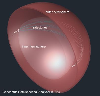
image that. We use a concentric hemispherical analyzer (CHA) because a CHA is
double focusing in both theta and phi (the two angular axes).
This illustration shows a set of trajectories focused at the center between two
(conducting) hemispheres. The outer hemisphere has a negative potential, and the
inner with a positive potential. Electrons with the right energy will form a focus at the
opposite side of the hemispheres where an aperture can be placed to allow only a
small range of energies to pass. We choose the pass energy by changing the
potential on the hemispheres.
created.
These photoelectrons have a range of energies from the true secondary distribution
at 1-2 eV up to the energy of the x-rays.
The photoelectrons are ejected into the magnetic field, and travel down the field
lines until they arrive at the field terminating aperture.
The angle which the electrons have after exiting the magnetic field depends not only
on their original position on the sample but also their kinetic energy, and the final
field just before the field terminating aperture.
The angular image at the field termination is therefore a confused superposition of
images at different energies.
We need to use an electron energy analyzer to extract one energy of interest and
image that. We use a concentric hemispherical analyzer (CHA) because a CHA is
double focusing in both theta and phi (the two angular axes).
This illustration shows a set of trajectories focused at the center between two
(conducting) hemispheres. The outer hemisphere has a negative potential, and the
inner with a positive potential. Electrons with the right energy will form a focus at the
opposite side of the hemispheres where an aperture can be placed to allow only a
small range of energies to pass. We choose the pass energy by changing the
potential on the hemispheres.
To get the angular image coming out of the magnetic field into the CHA we use a
transfer lens. This lens also gives some distance between any stray magnetic
fields and the electrostatic CHA.
In its simplest form the transfer lens is a three element cylindrical tube as shown
here. An accelerating voltage is put on the center element and the photoelectrons
are focused by the electrostatic fields between the cylinders, as shown in the
illustration.
The advantage of this arraignment is that a lens with more elements than shown
here allows us to change the angular magnification, and final energy of the
electrons entering the CHA.
Note, that an accelerating electrostatic lens has low aberration coefficients.

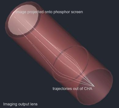
When the monochromatic photoelectron image leaves the CHA it must be then
focused onto a flat image plane (a phosphor plate) for image capture.
We again use an accelerating electrostatic cylindrical tube lens to focus the image
A simple two element lens acts like a telescope lens turning the monchromatic
angular image into a plane image.
The image is projected onto a phosphor screen, and the image is captured
outside of the vacuum chamber using a CCD camera.
The angular image out of the magnet must be energy analyzed and projected as a real x.y
image. We use a concentric hemispherical analyser with an input and output lens.
image. We use a concentric hemispherical analyser with an input and output lens.
More information on how it works

Early image of two superimposed grids.
Note high depth of focus
Note high depth of focus
| | | | | | | |
| How it works |